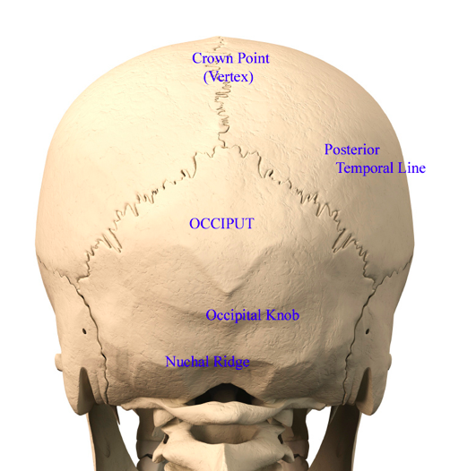
Back Of Skull Anatomy : The Human Skull Anatomical Chart - Anatomy Models and ... : The back muscles can be three types.. Most of the bones of the skull are held together by firm, immovable fibrous joints called sutures or synarthroses. These layers of back muscles help to mobilize and stabilize your trunk during your day to day activities. Spinal anatomy is a remarkable combination of strong bones, flexible ligaments and tendons, large muscles and highly sensitive nerves. The bones of the skull are highly irregular. Occipital bone anatomy the occipital bone is an unpaired bone, which covers the back of the head.
As these bones grow throughout fetal and childhood development, they begin to fuse together, forming a single skull. These layers of back muscles help to mobilize and stabilize your trunk during your day to day activities. May 31, 2021 · skull anatomy diagrams. Your back consists of three distinct layers of muscles, namely the superficial layer, the intermediate layer, and the deep layer. Intermediate back muscles and c.
Back of head skull anatomy Dr Barry Eppley Indianapolis ... from exploreplasticsurgery.com Your back consists of three distinct layers of muscles, namely the superficial layer, the intermediate layer, and the deep layer. Occipital bone anatomy the occipital bone is an unpaired bone, which covers the back of the head. The branching pattern of this artery forms readily visible grooves on the internal surface of the skull and these grooves can be traced back to their origin at the foramen spinosum. The back muscles can be three types. It contains the osteology, arthrology and myology of the spine and back. As these bones grow throughout fetal and childhood development, they begin to fuse together, forming a single skull. Sep 22, 2020 · this human anatomy module is composed of diagrams, illustrations and 3d views of the back, cervical, thoracic and lumbar spinal areas as well as the various vertebrae. Most of the bones of the skull are held together by firm, immovable fibrous joints called sutures or synarthroses.
The spine is made of 33 individual bones stacked one on top of the other.
As these bones grow throughout fetal and childhood development, they begin to fuse together, forming a single skull. The back muscles can be three types. It is designed to be incredibly strong, protecting the highly sensitive nerve roots, yet highly flexible, providing for mobility on many different planes. Deep back muscles superficial back muscles action movements of the shoulder. The branching pattern of this artery forms readily visible grooves on the internal surface of the skull and these grooves can be traced back to their origin at the foramen spinosum. The muscles of the back that work together to support the spine, help keep the body upright and allow twist and bend in many directions. May 31, 2021 · skull anatomy diagrams. May 19, 2021 · anatomy of back muscles. Anatomy of the spine overview. Occipital bone anatomy the occipital bone is an unpaired bone, which covers the back of the head. As mentioned, the skull is home to so many structures that the prospect of learning them all can seem very overwhelming. This spinal column provides the main support for your body, allowing you to stand upright, bend, and twist, while protecting the spinal cord from injury. These layers of back muscles help to mobilize and stabilize your trunk during your day to day activities.
During fetal development, the bones of the skull form within tough, fibrous membranes in a fetus' head. Mar 23, 2021 · in all, there are 22 bones comprising the entire skull, excluding the 3 pairs of ossicles located in the inner ear. Occipital bone anatomy the occipital bone is an unpaired bone, which covers the back of the head. These layers of back muscles help to mobilize and stabilize your trunk during your day to day activities. Most of the bones of the skull are held together by firm, immovable fibrous joints called sutures or synarthroses.
Posterior and lateral views of the skull: Anatomy | Kenhub from thumbor.kenhub.com During fetal development, the bones of the skull form within tough, fibrous membranes in a fetus' head. These layers of back muscles help to mobilize and stabilize your trunk during your day to day activities. This spinal column provides the main support for your body, allowing you to stand upright, bend, and twist, while protecting the spinal cord from injury. Anatomy of the spine overview. The back muscles can be three types. Deep back muscles superficial back muscles action movements of the shoulder. Sep 22, 2020 · this human anatomy module is composed of diagrams, illustrations and 3d views of the back, cervical, thoracic and lumbar spinal areas as well as the various vertebrae. Spinal anatomy is a remarkable combination of strong bones, flexible ligaments and tendons, large muscles and highly sensitive nerves.
These layers of back muscles help to mobilize and stabilize your trunk during your day to day activities.
Anatomy of the spine overview. Deep back muscles superficial back muscles action movements of the shoulder. During fetal development, the bones of the skull form within tough, fibrous membranes in a fetus' head. This spinal column provides the main support for your body, allowing you to stand upright, bend, and twist, while protecting the spinal cord from injury. The muscles of the back that work together to support the spine, help keep the body upright and allow twist and bend in many directions. The branching pattern of this artery forms readily visible grooves on the internal surface of the skull and these grooves can be traced back to their origin at the foramen spinosum. Mar 23, 2021 · in all, there are 22 bones comprising the entire skull, excluding the 3 pairs of ossicles located in the inner ear. As mentioned, the skull is home to so many structures that the prospect of learning them all can seem very overwhelming. Most of the bones of the skull are held together by firm, immovable fibrous joints called sutures or synarthroses. Intermediate back muscles and c. May 31, 2021 · skull anatomy diagrams. Spinal anatomy is a remarkable combination of strong bones, flexible ligaments and tendons, large muscles and highly sensitive nerves. Occipital bone anatomy the occipital bone is an unpaired bone, which covers the back of the head.
These layers of back muscles help to mobilize and stabilize your trunk during your day to day activities. Your back consists of three distinct layers of muscles, namely the superficial layer, the intermediate layer, and the deep layer. The branching pattern of this artery forms readily visible grooves on the internal surface of the skull and these grooves can be traced back to their origin at the foramen spinosum. As mentioned, the skull is home to so many structures that the prospect of learning them all can seem very overwhelming. During fetal development, the bones of the skull form within tough, fibrous membranes in a fetus' head.
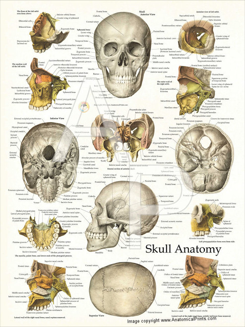
Skull Anatomy and Facial Structures Poster - Clinical ... from cdn1.bigcommerce.com Most of the bones of the skull are held together by firm, immovable fibrous joints called sutures or synarthroses. The muscles of the back that work together to support the spine, help keep the body upright and allow twist and bend in many directions. Sep 22, 2020 · this human anatomy module is composed of diagrams, illustrations and 3d views of the back, cervical, thoracic and lumbar spinal areas as well as the various vertebrae. The spine is made of 33 individual bones stacked one on top of the other. During fetal development, the bones of the skull form within tough, fibrous membranes in a fetus' head. The branching pattern of this artery forms readily visible grooves on the internal surface of the skull and these grooves can be traced back to their origin at the foramen spinosum. It contains the osteology, arthrology and myology of the spine and back. As these bones grow throughout fetal and childhood development, they begin to fuse together, forming a single skull.
Sep 22, 2020 · this human anatomy module is composed of diagrams, illustrations and 3d views of the back, cervical, thoracic and lumbar spinal areas as well as the various vertebrae.
Occipital bone anatomy the occipital bone is an unpaired bone, which covers the back of the head. It is designed to be incredibly strong, protecting the highly sensitive nerve roots, yet highly flexible, providing for mobility on many different planes. The spine is made of 33 individual bones stacked one on top of the other. Sep 22, 2020 · this human anatomy module is composed of diagrams, illustrations and 3d views of the back, cervical, thoracic and lumbar spinal areas as well as the various vertebrae. Spinal anatomy is a remarkable combination of strong bones, flexible ligaments and tendons, large muscles and highly sensitive nerves. Your back consists of three distinct layers of muscles, namely the superficial layer, the intermediate layer, and the deep layer. The branching pattern of this artery forms readily visible grooves on the internal surface of the skull and these grooves can be traced back to their origin at the foramen spinosum. May 31, 2021 · skull anatomy diagrams. The bones of the skull are highly irregular. During fetal development, the bones of the skull form within tough, fibrous membranes in a fetus' head. It contains the osteology, arthrology and myology of the spine and back. Deep back muscles superficial back muscles action movements of the shoulder. Most of the bones of the skull are held together by firm, immovable fibrous joints called sutures or synarthroses.


0 Komentar